Compact Bone Diagram Lacunae / Pin on A&P : Within these rings, are space called lacunae that contain osteocytes.
byAdmin-
0
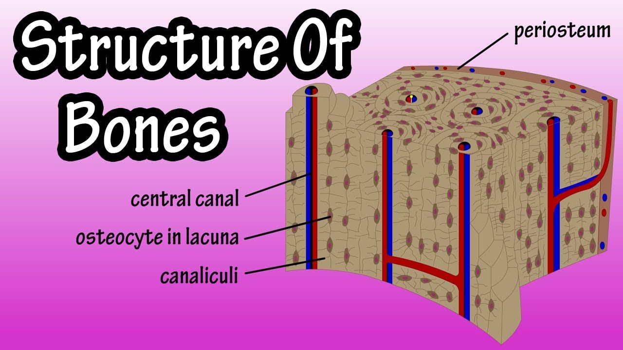
Compact Bone Diagram Lacunae / Pin on A&P : Within these rings, are space called lacunae that contain osteocytes.. Bone canaliculi are microscopic canals between the lacunae of ossified bone. The osteocytes are sitting in the lacunae and the canals are canaliculi, which interconnect the lacunae with the major vessels. Like compact bone, spongy bone, also known as cancellous bone, contains osteocytes housed in lacunae, but diagram of blood and nerve supply to bone. Start studying structure of compact bone. Lacunae is really just an empty space for osteocytes or bone cells and these osteocytes have these long cellular processes that branch through the canaliculi to contact other osteocytes via gap junctions which allow these cells to communicate with each other and exchange nutrients and and signals with.
It is a bone is one of two kinds of bone tissue that can be found in the body of a only tiny spaces (lacunae) are left which that contain the bone cells or osteocytes. The lacunae of bones consist of canaliculi between osteocytes. Compact bone is arranged in concentric ring structures called osteons (haversian canal system) in the center of each ring is a structure called a haversian canal. Compact bone is sometimes called cortical bone. To know the structures of a synovial joint and a symphysis joint (intervertebral disc).
Compact Bone Diagram Pearson : Medullary Cavity Wikipedia ... from i.pinimg.com The small open spaces created in the lamellae by the osteocytes are called lacunae. Histologically, two important bone types are compact bone and spongy bone. 6 compact bone vs spongy bone. Like compact bone, spongy bone, also known as cancellous bone, contains osteocytes housed in lacunae, but they are not arranged in concentric circles. Compact bone is also called cortical bone whereas the spongy bone has interstitial lamellae occupy the space between adjacent concentric lamellae. The series of diagrams below represent the microscopic structure of compact bone tissue. Compact bone contains cylinders of calcified bone known as osteons or haversian systems. Compact bone, concentric lamellae, haversion system, haversion canal, osteocytes.
The lacunae of bones consist of canaliculi between osteocytes.
In histology, a lacuna is a small space, containing an osteocyte in bone, or chondrocyte in cartilage. Thin layer of reticular ct lining internal marrow cavity. Each haversian canal is surrounded by concentric rings of compact bone called lamellae. The small open spaces created in the lamellae by the osteocytes are called lacunae. The basic units of compact bone are called osteons or haversian systems. The proportion of bone tissue occupied by osteocyte lacunae (% c/t ) appears to follow at different levels of long bones, the same pattern recorded for the data of bone turnover rate, by the tetracycline labeling technique: The lacunae are situated between the lamellae, and consist of a number of oblong spaces. You should include the histology of compact bone slides with diagram as well into your article. Histologically, two important bone types are compact bone and spongy bone. Compact bone is also called cortical bone whereas the spongy bone has interstitial lamellae occupy the space between adjacent concentric lamellae. Learn vocabulary, terms and more with flashcards, games and other study tools. In three dimensions an osteon is cylindrical in shape. Compact bone consists of closely packed osteons or haversian systems.
To know the architecture of compact and spongy (cancellous) bone. You should include the histology of compact bone slides with diagram as well into your article. In three dimensions an osteon is cylindrical in shape. Bone osseous tissue labeled cancellous bone structure spongy bone diagram compact bone connective tissue vascular lacunae compact bone 400x haversian system bone osteocyte function osteoblast osteocyte osteoclast bone cell. Like compact bone, spongy bone, also known as cancellous bone, contains osteocytes housed in lacunae, but they are not arranged in concentric circles.
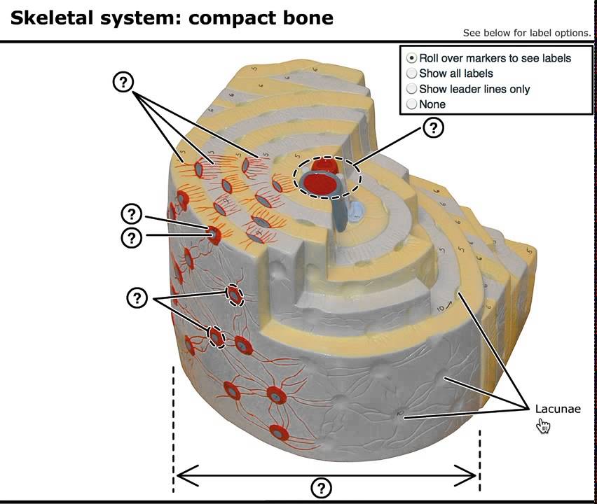
Structure Of Bone Tissue - Bone Structure Anatomy ... from i.ytimg.com A diagram of a section of compact bone showing haversian canals. To know the architecture of compact and spongy (cancellous) bone. Compact bone is sometimes called cortical bone. Thin layer of reticular ct lining internal marrow cavity. Compact bone, concentric lamellae, haversion system, haversion canal, osteocytes. The outlined area is a cross section of an osteon of compact bone. The osteon consists of a central canal called the osteonic (haversian) canal, which is surrounded by concentric rings (lamellae) of matrix. In histology, a lacuna is a small space, containing an osteocyte in bone, or chondrocyte in cartilage.
The two types of bones are compact bones and spongy bones.
Compact bone is arranged in concentric ring structures called osteons (haversian canal system) in the center of each ring is a structure called a haversian canal. In living bone, osteocytic lacunae contain osteocytes and they are the. Start studying structure of compact bone. In an ordinary microscopic section, viewed by transmitted light, they appear as fusiform opaque spots. It can be remodeled all throughout life to withstand stress. The three types of cartilages are image courtesy: Learn vocabulary, terms and more with flashcards, games and other study tools. The proportion of bone tissue occupied by osteocyte lacunae (% c/t ) appears to follow at different levels of long bones, the same pattern recorded for the data of bone turnover rate, by the tetracycline labeling technique: To know the structures of a synovial joint and a symphysis joint (intervertebral disc). Find stockbilleder af anatomy compact bone i hd og millionvis af andre royaltyfri stockbilleder, illustrationer og vektorer i shutterstocks samling. Compact bone contains cylinders of calcified bone known as osteons or haversian systems. Around the haversian canal, rings of bone tissue are found called lamellae. Rather, the osteocytes containing lacunae are arranged in a.
It can be remodeled all throughout life to withstand stress. Once the osteoid is mineralized, the precursor cells get surrounded by organic intracellular substances called lacunae to become fully developed and matured into osteocytes. The three dimensional functional units within compact bone are called osteons. You should include the histology of compact bone slides with diagram as well into your article. Bone canaliculi are microscopic canals between the lacunae of ossified bone.
In The Diagram Where Is The Osteon - Atkinsjewelry from i.ytimg.com Compact bone tissue diagram quizlet. Once the osteoid is mineralized, the precursor cells get surrounded by organic intracellular substances called lacunae to become fully developed and matured into osteocytes. The outlined area is a cross section of an osteon of compact bone. You should include the histology of compact bone slides with diagram as well into your article. Tusindvis af nye billeder af høj kvalitet tilføjes hver dag. Like compact bone, spongy bone, also known as cancellous bone, contains osteocytes housed in lacunae, but they are not arranged in concentric circles. Unit 4 (anat1) (bone, cartilage, connective tissue, connective tissue proper, bone tissue, ossification). (singular = lacuna) spaces in a bone that house an osteocyte.
Around the haversian canal, rings of bone tissue are found called lamellae.
Thin layer of reticular ct lining internal marrow cavity. The walls of the diaphysis are composed of dense and hard compact bone. Like compact bone, spongy bone, also known as cancellous bone, contains osteocytes housed in lacunae, but they are not arranged in concentric circles. Osteocytes occupy spaces (lacunae) in the bone matrix. Compact bone is also called cortical bone whereas the spongy bone has interstitial lamellae occupy the space between adjacent concentric lamellae. The outlined area is a cross section of an osteon of compact bone. Within these rings, are space called lacunae that contain osteocytes. (singular = lacuna) spaces in a bone that house an osteocyte. In living bone, osteocytic lacunae contain osteocytes and they are the. The three types of cartilages are image courtesy: The lacunae are situated between the lamellae, and consist of a number of oblong spaces. Compact bone consists of closely packed osteons or haversian systems. The musculoskeletal system is comprised of bones and connective tissue structures, such as cartilage, ligaments, and tendons.
Lacunae actually means, a lake in latin compact bone diagram. It is a bone is one of two kinds of bone tissue that can be found in the body of a only tiny spaces (lacunae) are left which that contain the bone cells or osteocytes.